Fraud Blocker Skip to main content

Pillow > Science > SEM

SEM Pillow Collection (#4)

1,342 Pillows

Background imageSEM Pillow Collection: Globorotalia scitula, foraminifera fossil

Globorotalia scitula, foraminifera fossil
Scanning electron microscope (SEM) image showing a fossilised planktonic species of foraminifera

Background imageSEM Pillow Collection: Scyliorhinus canicula, dogfish

Scyliorhinus canicula, dogfish
Scanning electron microscope (SEM) image of the scales of a dogfish (x 40)

Background imageSEM Pillow Collection: Needle playing a record, SEM

Needle playing a record, SEM
Needle playing a record. Coloured scanning electron micrograph (SEM) of the needle (stylus) of a record player in a groove on a record. A record is used to store sound

Background imageSEM Pillow Collection: Ragworm mouth, SEM

Ragworm mouth, SEM
Ragworm mouth. Coloured scanning electron micrograph (SEM) of the mouth of a ragworm (Nereis sp.). Four teeth (cream) are seen around the opening. These worms are used commercially as fishing bait

Background imageSEM Pillow Collection: Nematode worm, SEM

Nematode worm, SEM
Nematode worm, coloured scanning electron micrograph (SEM). Nematode worms are small worms that are unsegmented. Depending on the species, a nematode worm may be parasitic or free-living

Background imageSEM Pillow Collection: SEM of bed bugs

SEM of bed bugs
Bed bugs mating. Coloured scanning electron micrograph (SEM) of a pair of bed bugs mating. Bed bugs (Cimex lectularius)are blood sucking insects, so called because they live in the beds of humans

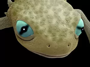
Background imageSEM Pillow Collection: Head of a young newt, SEM

Head of a young newt, SEM
Head of a young newt. Scanning electron micrograph (SEM) of a juvenile (immature) newt. Although this newt is in the last stages of development

Background imageSEM Pillow Collection: Blowfly laying eggs, SEM

Blowfly laying eggs, SEM
Blowfly laying eggs. Coloured scanning electron micrograph (SEM) of a female Lucilia sp. blowfly laying her eggs (lower left). A blowfly lays its eggs on dead bodies

Background imageSEM Pillow Collection: Coloured SEM of egg & sperm during fertilisation

Coloured SEM of egg & sperm during fertilisation
Fertilisation. Coloured Scanning Electron Micro- graph (SEM) of a human egg (ovum) being penetrated by sperm during fertilisation. Sperm are coloured yellow, each with a rounded head and a long tail

Background imageSEM Pillow Collection: Kidney glomeruli, SEM

Kidney glomeruli, SEM
Kidney glomeruli, coloured scanning electron micrograph (SEM). The cells that usually surround the glomeruli have been removed to reveal the coiled capillaries at the core of the glomeruli

Background imageSEM Pillow Collection: Red blood cells, SEM

Red blood cells, SEM
Red blood cells, coloured scanning electron micrograph (SEM). Red blood cells (erythrocytes) are biconcave, giving them a large surface area for gas exchange, and highly elastic

Background imageSEM Pillow Collection: Phagocytosis of fungal spores, SEM

Phagocytosis of fungal spores, SEM
Phagocytosis of a fungus spore. Coloured scanning electron micrograph (SEM) of a neutrophil (blue) phagocytosing (engulfing and destroying) spores from the fungus Aspergillus fumigatus (red)

Background imageSEM Pillow Collection: Used dental floss, SEM

Used dental floss, SEM
Used dental floss. Coloured scanning electron micrograph (SEM) of dental plaque on strands of used dental floss. Plaque consists of a film of bacteria embedded in a glycoprotein matrix

Background imageSEM Pillow Collection: False-colour SEM showing two renal glomeruli

False-colour SEM showing two renal glomeruli
False-colour scanning electron micrograph of a section of kidney cortex, showing two glomeruli (red & green). A glomerulus is a tiny knot of blood capillaries which lies cupped within a Bowmans

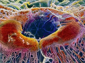
Background imageSEM Pillow Collection: False-colour SEM of retina featuring central fovea

False-colour SEM of retina featuring central fovea
Foveal retina. False-colour scanning electron micrograph of the human retina featuring the central fovea, a crater-like depression in the photosensitive layer of the eye

Background imageSEM Pillow Collection: Coloured SEM of a section through nerve fibres

Coloured SEM of a section through nerve fibres
Nerve fibres. Coloured scanning electron micrograph (SEM) of a section through bundles of nerve fibres (red). Each is within a sheath, or perineurium (blue)

Background imageSEM Pillow Collection: Neutrophil cell trapping bacteria, SEM

Neutrophil cell trapping bacteria, SEM
Neutrophil cell trapping bacteria. Coloured scanning electron micrograph (SEM) of bacteria (rod-shaped) being trapped by a neutrophil cell

Background imageSEM Pillow Collection: Cauliflower head, SEM

Cauliflower head, SEM
Cauliflower. Coloured scanning electron micrograph (SEM) of the head of a cauliflower (Brassica oleracea botrytis). The arrangement of florets around the head is according to a Fibonacci series

Background imageSEM Pillow Collection: Fossilised diatoms, SEM

Fossilised diatoms, SEM

Background imageSEM Pillow Collection: Kidney glomerulus, SEM

Kidney glomerulus, SEM
Kidney glomerulus, coloured scanning electron micrograph (SEM). Podocyte cells, which make up the surface of the glomerulus, are yellow. Magnification: x650 when printed at 10 centimetres wide

Background imageSEM Pillow Collection: Carpet beetle larva, SEM

Carpet beetle larva, SEM
Carpet beetle larva. Coloured scanning electron micrograph (SEM) of a carpet beetle (Anthrenus sp.) larva amongst the fibres of a wool carpet

Background imageSEM Pillow Collection: Macrophage cells, SEM

Macrophage cells, SEM
Macrophage cells. Coloured scanning electron micrograph (SEM) of macrophage white blood cells. Macrophages are cells of the bodys immune system

Background imageSEM Pillow Collection: Ovarian follicle, SEM

Ovarian follicle, SEM
Ovarian follicle. Coloured scanning electron micrograph (SEM) of a fracture through a secondary follicle in the ovary. The oocyte (developing egg) is orange and its central nucleus is darker orange

Background imageSEM Pillow Collection: Dinoflagellate plankton, SEM

Dinoflagellate plankton, SEM
Dinoflagellate plankton. Coloured scanning electron micrograph (SEM) of the calcareous (calcium-based) test (shell) of a dinoflagellate Calciodinellum sp

Background imageSEM Pillow Collection: Spiny spider, SEM

Spiny spider, SEM
Spiny spider. Coloured scanning electron micrograph (SEM) of a spiny spider (Gasteracantha sp.), showing its characteristic spines (red) and large mouthparts (grey, centre left)

Background imageSEM Pillow Collection: Trachea lining, SEM

Trachea lining, SEM
Trachea lining, coloured scanning electron micrograph (SEM). The tracheal lining contains mucus-secreting goblet cells and epithelial cells bearing hair-like cilia (blue)

Background imageSEM Pillow Collection: Dandelion pollen grain, SEM

Dandelion pollen grain, SEM
Dandelion (Taraxacum officinale) pollen grain, coloured scanning electron micrograph (SEM). Pollen grains are reproductive structures produced by the male parts of flowering plants

Background imageSEM Pillow Collection: Golgi apparatus, SEM

Golgi apparatus, SEM
Golgi apparatus, coloured scanning electron micrograph (SEM). Section through a liver cell showing its Golgi apparatus (grey), a membrane-bound organelle that modifies and packages proteins

Background imageSEM Pillow Collection: Goosegrass fruits, SEM

Goosegrass fruits, SEM
Goosegrass fruits. Coloured scanning electron micrograph (SEM) of the hooked fruits of a goosegrass plant (Galium aparine)

Background imageSEM Pillow Collection: Synapse nerve junctions, SEM

Synapse nerve junctions, SEM
Synapse nerve junctions. Coloured scanning electron micrograph (SEM) of nerve cells showing the synapses (junctions, bulges) between them

Background imageSEM Pillow Collection: Silver birch twig, SEM

Silver birch twig, SEM
Silver birch twig. Coloured scanning electron micrograph (SEM) of a section through a silver birch (Betula pendula) twig. At the centre of the twig (top centre left) is the pith

Background imageSEM Pillow Collection: Morning glory pollen, SEM

Morning glory pollen, SEM
Morning glory pollen grains. Coloured scanning electron micrograph (SEM) of pollen grains (orange balls) on the pistil (female reproductive part) of a morning glory flower (Ipomoea sp.)

Background imageSEM Pillow Collection: Forget-me-not flower, SEM

Forget-me-not flower, SEM
Forget-me-not flower. Coloured scanning electron micrograph (SEM) of a forget-me-not (Myosotis arvensis) flower. The petals (blue) are covered by numerous projections called papillae

Background imageSEM Pillow Collection: Pollen on a bee leg, SEM

Pollen on a bee leg, SEM
Pollen on a bee leg. Coloured scanning electron micrograph (SEM) of pollen grains (pale purple) on the leg of a bee (superfamily Apoidea). Pollen grains are the male sex cells of a flowering plant

Background imageSEM Pillow Collection: Pseudomonas aeruginosa bacteria, SEM

Pseudomonas aeruginosa bacteria, SEM
Pseudomonas aeruginosa bacteria, coloured scanning electron micrograph (SEM). These Gram-negative rod-shaped bacteria are found in soil and water, and as normal flora in the human intestine

Background imageSEM Pillow Collection: Scanning Electron Micrograph (SEM): Common House Spider - male, Magnification x (A4 size: 29)

Scanning Electron Micrograph (SEM): Common House Spider - male, Magnification x (A4 size: 29. 7 cm width)
LRDS-119 Common House Spider - male Scanning Electron Micrograph (SEM) Tegenaria domestica Magnification x (A4 size: 29.7 cm width) Coloured by hand to enhance natural features

Background imageSEM Pillow Collection: Scanning Electron Micrograph (SEM): Jumping Spider, Magnification x 75 (A4 size: 29. 7 cm width)

Scanning Electron Micrograph (SEM): Jumping Spider, Magnification x 75 (A4 size: 29. 7 cm width)
LRDS-103 Jumping Spider Scanning Electron Micrograph (SEM) Portia sp. Magnification x 75 (A4 size: 29.7 cm width) Coloured by hand to enhance natural features

Background imageSEM Pillow Collection: Scanning Electron Micrograph (SEM): Termite (worker) - Magnification x 100 (if print A4 size: 29)

Scanning Electron Micrograph (SEM): Termite (worker) - Magnification x 100 (if print A4 size: 29. 7 cm wide)
LRDS-410 Scanning Electron Micrograph (SEM): Termite (worker) Magnification x 100 (if print A4 size: 29.7 cm wide) Reticulitermes lucifugus David Spears (Last Refuge)

Background imageSEM Pillow Collection: Fly on sugar, SEM

Fly on sugar, SEM
Fly on sugar, coloured scanning electron micrograph (SEM). Flies (order Diptera) are insects that can spread disease when they feed on human food

Background imageSEM Pillow Collection: SEM of a hooks and loops fastener

SEM of a hooks and loops fastener
Coloured scanning electron micrograph (SEM)of a hooks and loops fastener. It is a two-sheet material that is used as a reversible fastener on clothing and fabrics

Background imageSEM Pillow Collection: Salt grain and peppercorn, SEM

Salt grain and peppercorn, SEM
Salt and peppercorn. Coloured scanning electron micrograph (SEM) of a salt grain (blue) and a peppercorn (brown). Salt (sodium chloride) is a crystalline compound

Background imageSEM Pillow Collection: Osteocyte bone cell, SEM

Osteocyte bone cell, SEM
Osteocyte bone cell. Coloured scanning electron micrograph (SEM) of an osteocyte bone cell (blue) surrounded by bone tissue (grey)

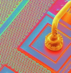
Background imageSEM Pillow Collection: Microchip connecting wire, SEM

Microchip connecting wire, SEM
Micro-wire. Coloured scanning electron micrograph (SEM) of a micro-wire (yellow, often made of gold) connecting to the surface of a microchip

Background imageSEM Pillow Collection: Water bear, SEM C016 / 9084

Water bear, SEM C016 / 9084
Water bear (Echiniscus granulatus). Coloured scanning electron micrograph (SEM) showing the underside of a water bear, or tardigrade (phylum Tardigrada)

Background imageSEM Pillow Collection: Suckling clover (Trifolium dubium), SEM C015 / 9231

Suckling clover (Trifolium dubium), SEM C015 / 9231
Suckling clover (Trifolium dubium) flower, coloured scanning electron micrograph (SEM)

Background imageSEM Pillow Collection: Aspergillus

Aspergillus
An SEM image of aspergillus in spore production (x 815 on a standard 9 cm wide print). The moulds are common in the northern hemisphere and some cause disease in humans and animals

Background imageSEM Pillow Collection: Caterpillar egg

Caterpillar egg
Scanning electron microscope image of a caterpillar egg (x 90), the caterpillar emerges by chewing through the shell (x 350)

Background imageSEM Pillow Collection: Blackfly antenna

Blackfly antenna
Scanning electron microscope image of a blackfly antenna (x 350). These long sensory organs feel and taste objects as well as sensing vibrations and smells (x 1.1K)


All Professionally Made to Order for Quick Shipping


-

Why Choose Us?

We are a leading provider of Art Prints and Photo Gifts since 2003, working in partnership with a range of Sporting Clubs, Charities, Museums and Picture Libraries. A large share of profits from any of their images will go directly towards supporting that charity or club. Our archive of images is carefully curated to bring you a wide range of subjects, including landscapes, wildlife, architecture, and more. We ship from our partner labs in the UK, USA, EU (Netherlands) and Australia.
+

How do I place an order?

Ordering is quick & easy - Just follow 5 Simple Steps:
  1. Find your image: Use our search box or browse our online photo Collections to find the image you want.

  2. Choose your print format: Select your desired product and add it to your cart.

  3. Enter your details: If you're a returning customer, simply enter your email address and password, and we'll fill in your billing and shipping address details. All personal details are held securely and are fully GDPR compliant. As standard, we remove all Personally Identifiable Information after 12 months.

  4. Pay for your purchase: We use state-of-the-art security for online shopping and do not have access to your card details.

  5. Sit back and relax: We'll email you confirmation of your order and when it's dispatched. Registered customers can also track orders in the 'My Account' area.

+

How do I pay for an order?

You can pay for your order with most of the major credit and debit cards, or PayPal. For added security, major financial institutions process payment details separately and securely on our behalf. We do not have access to your online payment card details. Online payments are preferred however we do take phone orders. For UK customers only we accept cheques issued against a UK bank.
+

Is my data safe?

We take data security very seriously. We do not have access to your full card details and all payments are requested over a fully secure connection. Additionally, we fully comply with current European and GDPR legislation, and automatically remove all personal data after at most 12 months (unless you have an account with us and have signed in recently). We also have a strict opt-in policy and would never sell your personal details. Your data is only used to fulfill your order promptly and efficiently.
+

Is wall art ready to hang?

For quick and easy installation all wall art, including framed prints, canvas prints and metal prints are supplied with a ready to hang solution on the back. Generally, saw tooth hangers are applied as they allow wall art to hang flush against the wall. The serrated edge of the hanger prevents the frame from shifting or tilting when hung.
+

Are the photo prints fade resistant?

Yes, we use archival quality photo paper photographic paper for vivid reproduction Prints are an accurate representations of the original artwork, which is preserved for artistic character and authenticity. We guarantee they match previews shown on our web site