Fraud Blocker Skip to main content

Photographic Print > Science > SEM

SEM Photographic Print Collection (#28)

1,342 Photographic Prints

Background imageSEM Photographic Print Collection: Turtle ants head, SEM

Turtle ants head, SEM
Turtle ants head. Coloured scanning electron micrograph (SEM) of the head of a soldier turtle ant (Cephalotes sp.) from the Amazonian rainforest. One of its compound eyes (green) is at left

Background imageSEM Photographic Print Collection: False-colour SEM of the head of a housefly

False-colour SEM of the head of a housefly
Coloured scanning electron micrograph of the head of a female housefly, Musca domestica. The head is dominated by the large compound eyes, here tinted yellow

Background imageSEM Photographic Print Collection: Female mosquito head, SEM

Female mosquito head, SEM
Female mosquito head. Coloured scanning electron micrograph (SEM) of the head of a female mosquito (family Culicidae). The mosquitos head is dominated by its large compound eyes (pink)

Background imageSEM Photographic Print Collection: Coloured SEM of a human hair on the skin

Coloured SEM of a human hair on the skin
Hair on skin. Coloured Scanning Electron Micro- graph (SEM) of a hair on the surface of human skin. At centre is a shaft of hair (dark grey) which has emerged from a follicle in the skin

Background imageSEM Photographic Print Collection: Eyelash hairs, SEM

Eyelash hairs, SEM
Eyelash hairs and skin. Coloured scanning electron micrograph (SEM) of eyelash hairs growing from the surface of human skin

Background imageSEM Photographic Print Collection: Coloured SEM of a section through a hair follicle

Coloured SEM of a section through a hair follicle
Hair follicle. Coloured scanning electron micrograph (SEM) of a cross-section through a hair follicle below the skin surface

Background imageSEM Photographic Print Collection: False-colour SEM of embryo at the morula stage

False-colour SEM of embryo at the morula stage
Embryo development. False-colour scanning electron micrograph of an embryo at the early stage known as the morula. The egg reaches this phase about 4 days after fertilisation after a series of

Background imageSEM Photographic Print Collection: Alveolus in human lung

Alveolus in human lung
Alveolus. False-colour scanning electron micrograph (SEM) of an alveolus (blue cavity), one of the microscopic air sacs of the lungs

Background imageSEM Photographic Print Collection: Bone tissue

Bone tissue. Coloured scanning electron micrograph (SEM) of cancellous (spongy) bone. Bone tissue can be either cortical (compact) or cancellous

Background imageSEM Photographic Print Collection: Metal shavings

Metal shavings. Coloured scanning electron micrograph (SEM) of metal shavings taken from a key cutter s

Background imageSEM Photographic Print Collection: Allergens in trachea

Allergens in trachea. Coloured scanning electron micrograph (SEM) of the surface of the trachea (windpipe) with a number of pollen grains (yellow) attached

Background imageSEM Photographic Print Collection: Komodo dragon tooth, SEM

Komodo dragon tooth, SEM
Komodo dragon tooth, coloured scanning electron micrograph (SEM). This curved serrated tooth is designed to slice, cut and tear meat, but not to chew it

Background imageSEM Photographic Print Collection: False-colour SEM of beard hair cut by razor blade

False-colour SEM of beard hair cut by razor blade

Background imageSEM Photographic Print Collection: Trachea, SEM

Trachea, SEM
Trachea. Coloured scanning electron micrograph (SEM) of the lining of the trachea (windpipe). The trachea links the larynx (voice box) to the lungs

Background imageSEM Photographic Print Collection: SEM of rod & cone cell of the retina

SEM of rod & cone cell of the retina
False-colour scanning electron micrograph (SEM) of rod and cone cells of the eye retina. Cone cells (blue) and more numerous rod cells (pink, purple) are specialized light-sensitive cells

Background imageSEM Photographic Print Collection: Blood platelets

Blood platelets. Coloured scanning electron micro- graph (SEM) of activated blood platelets. Platelet cells (thrombocytes) are formed in the bone marrow

Background imageSEM Photographic Print Collection: False-colour SEM of blood cells on endocardium

False-colour SEM of blood cells on endocardium
Red Blood cells inside the heart: false-colour scanning electron micrograph (SEM) of red & white blood cells on the endocardium, the lining of the heart

Background imageSEM Photographic Print Collection: Osteoblast bone cell, SEM

Osteoblast bone cell, SEM
Osteoblast bone cell. Coloured scanning electron micrograph (SEM) of an osteoblast (centre) in bone tissue. Osteoblasts are primarily found in regions of new bone growth

Background imageSEM Photographic Print Collection: Ant, SEM

Ant, SEM
Ant (family Formicidae), coloured scanning electron micrograph (SEM). One of its compound eyes (black) is seen, as well as two long antennae that are mounted on the head between the eyes

Background imageSEM Photographic Print Collection: Mitochondria, SEM

Mitochondria, SEM
Mitochondria. Coloured scanning electron micrograph (SEM) of mitochondria (red) in a kidney cell. Mitochondria are a type of organelle found in the cytoplasm of eukaryotic cells

Background imageSEM Photographic Print Collection: Flakes of dead skin, SEM

Flakes of dead skin, SEM
Flakes of dead skin. Coloured scanning electron micrograph (SEM) of dead skin from the scalp (dandruff), a condition where there is an excessive shedding of dead skin cells

Background imageSEM Photographic Print Collection: Organ of Corti, inner ear, SEM

Organ of Corti, inner ear, SEM
Organ of Corti. Coloured scanning electron micrograph (SEM) of the organ of Corti from the cochlea of the inner ear. The bony capsule that encloses the organ has been removed to reveal the organs

Background imageSEM Photographic Print Collection: Chickweed pollen grain, SEM

Chickweed pollen grain, SEM
Chickweed pollen grain. Coloured scanning electron micrograph (SEM) of a pollen grain (green) from a chickweed flower (Stellaria media)

Background imageSEM Photographic Print Collection: Honey bee head, SEM

Honey bee head, SEM
Honey bee head. Coloured scanning electron micrograph (SEM) of the head of a honey bee (Apis sp.). The compound eyes (red) are either side of the antennae

Background imageSEM Photographic Print Collection: Lymphocytes in hair follicle, SEM

Lymphocytes in hair follicle, SEM
Lymphocytes in hair follicle, coloured scanning electron micrograph (SEM). Large numbers of lymphocytes (yellow) are seen in inflamed and necrotic tissue at the base of hair shafts (red)

Background imageSEM Photographic Print Collection: Bee foot claws, SEM

Bee foot claws, SEM
Bee foot claws, coloured scanning electron micrograph (SEM). These claws, on a bees fore legs, are used to comb pollen from the body and transfer it to the pollen baskets on the bees hind legs

Background imageSEM Photographic Print Collection: Bacillus subtilis bacteria, SEM

Bacillus subtilis bacteria, SEM
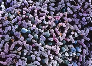
Bacillus subtilis. Coloured scanning electron micrograph (SEM) of Bacillus subtilis bacteria. B. subtilis, or hay bacillus, is an aerobic, gram-positive bacillus(rod-shaped bacterium)

Background imageSEM Photographic Print Collection: Lactobacillus bacteria, SEM

Lactobacillus bacteria, SEM
Lactobacillus sp. bacteria, coloured scanning electron micrograph (SEM). Lactobacillus bacteria are probiotic bacteria, which are useful for human and animal health as they produce bacteriocins

Background imageSEM Photographic Print Collection: Starch grains, SEM

Starch grains, SEM
Starch grains. Coloured scanning electron micrograph (SEM) of starch grains (orange) in a black-eyed pea (Vigna sp.). Starch is synthesised from sucrose

Background imageSEM Photographic Print Collection: Coloured SEM of Clostridium botulinum

Coloured SEM of Clostridium botulinum
Food poisoning bacterium. Coloured scanning electron micrograph of the Gram-positive anaerobic bacteria Clostridium botulinum, responsible for serious food poisoning in humans. C

Background imageSEM Photographic Print Collection: Root nodule

Root nodule. Coloured scanning electron micrograph (SEM) of a root nodule on a pea plant (Pisum sativum) caused by the nitrogen-fixing soil bacteria Rhizobium leguminosarum

Background imageSEM Photographic Print Collection: SEM of yeast cells

SEM of yeast cells
Yeast. Coloured scanning electron micrograph (SEM) of yeast cells, Saccharomyces cerevisiae, commonly known as Bakers or Brewers yeast, growing on potato dextrose agar

Background imageSEM Photographic Print Collection: Fennel stem, SEM

Fennel stem, SEM
Fennel stem. Coloured scanning electron micrograph (SEM) of a cluster of fennel (Foeniculum vulgare) flowers on a stem, seen from below. Magnification unknown

Background imageSEM Photographic Print Collection: Argan trees (Argania spinosa)

Argan trees (Argania spinosa) This tree is endemic to the Sous valley in Morocco. It is cultivated for the oil (argan oil) that is found in the fruit

Background imageSEM Photographic Print Collection: Marigold pollen grain, SEM

Marigold pollen grain, SEM
Marigold pollen grain. Coloured scanning electron micrograph (SEM) of a pollen grain from a marigold plant (family Compositae)

Background imageSEM Photographic Print Collection: Common wasp, SEM

Common wasp, SEM
Common wasp (Vespula vulgaris), coloured scanning electron micrograph (SEM). These wasps are social insects. They live in underground nests of up to around 10, 000 workers

Background imageSEM Photographic Print Collection: Bamboo shoot weevil, SEM

Bamboo shoot weevil, SEM
Bamboo shoot weevil (Otidognathus davidis), coloured scanning electron micrograph (SEM). This insect is a pest of bamboo shoots, and is widespread in the Far East

Background imageSEM Photographic Print Collection: Fungal spores, SEM

Fungal spores, SEM
Fungal spores. Coloured scanning electron micrograph (SEM) of fungal spores on a fruiting body of an Aspergillus sp. fungus. The name for such a fruiting body is a conidiophore

Background imageSEM Photographic Print Collection: Scanning Electron Micrograph (SEM): Termite (worker), ; Magnification x 65 (if print A4 size: 29)

Scanning Electron Micrograph (SEM): Termite (worker), ; Magnification x 65 (if print A4 size: 29. 7 cm wide)
LRDS-411 Scanning Electron Micrograph (SEM): Termite (worker), Magnification x 65 (if print A4 size: 29.7 cm wide) Reticulitermes lucifugus David Spears (Last Refuge)

Background imageSEM Photographic Print Collection: Scanning Electron Micrograph (SEM): Mosquito, Magnification x 35 (A4 size: 29. 7 cm width)

Scanning Electron Micrograph (SEM): Mosquito, Magnification x 35 (A4 size: 29. 7 cm width)
LRDS-84 Mosquito Scanning Electron Micrograph (SEM) Culex pipiens Magnification x 35 (A4 size: 29.7 cm width) Coloured by hand to enhance natural features

Background imageSEM Photographic Print Collection: Scanning Electron Micrograph (SEM): Flour Mite, Magnification x 500 (A4 size: 29. 7 cm width)

Scanning Electron Micrograph (SEM): Flour Mite, Magnification x 500 (A4 size: 29. 7 cm width)
LRDS-58 Flour Mite Scanning Electron Micrograph (SEM) Acarus siro Magnification x 500 (A4 size: 29.7 cm width) Coloured by hand to enhance natural features

Background imageSEM Photographic Print Collection: Scanning Electron Micrograph (SEM): Cat Flea, Magnification x250 (A4 size: 29. 7 cm width)

Scanning Electron Micrograph (SEM): Cat Flea, Magnification x250 (A4 size: 29. 7 cm width)
LRDS-54 Cat Flea Scanning Electron Micrograph (SEM) Ctenocephalides felis Magnification x250 (A4 size: 29.7 cm width) Coloured by hand to enhance natural features

Background imageSEM Photographic Print Collection: Scanning Electron Micrograph (SEM): Chigoe Flea, Magnification x 200 (A4 size: 29. 7 cm width)

Scanning Electron Micrograph (SEM): Chigoe Flea, Magnification x 200 (A4 size: 29. 7 cm width)
LRDS-137 Chigoe Flea Scanning Electron Micrograph (SEM) Tunga penetrans Magnification x 200 (A4 size: 29.7 cm width) Coloured by hand to enhance natural features

Background imageSEM Photographic Print Collection: Scanning Electron Micrograph (SEM): Springtail, Order Collembola; Magnification x 150 (A4 size)

Scanning Electron Micrograph (SEM): Springtail, Order Collembola; Magnification x 150 (A4 size: 29. 7 cm width)
LRDS-122 Springtail, Order Collembola Scanning Electron Micrograph (SEM) Magnification x 150 (A4 size: 29.7 cm width) Coloured by hand to enhance natural features

Background imageSEM Photographic Print Collection: Mehpeyker Sultan with her court servants, 1647. Artist: Anonymous

Mehpeyker Sultan with her court servants, 1647. Artist: Anonymous
Mehpeyker Sultan with her court servants, 1647. From a private collection


All Professionally Made to Order for Quick Shipping


-

Why Choose Us?

We are a leading provider of Art Prints and Photo Gifts since 2003, working in partnership with a range of Sporting Clubs, Charities, Museums and Picture Libraries. A large share of profits from any of their images will go directly towards supporting that charity or club. Our archive of images is carefully curated to bring you a wide range of subjects, including landscapes, wildlife, architecture, and more. We ship from our partner labs in the UK, USA, EU (Netherlands) and Australia.
+

How do I place an order?

Ordering is quick & easy - Just follow 5 Simple Steps:
  1. Find your image: Use our search box or browse our online photo Collections to find the image you want.

  2. Choose your print format: Select your desired product and add it to your cart.

  3. Enter your details: If you're a returning customer, simply enter your email address and password, and we'll fill in your billing and shipping address details. All personal details are held securely and are fully GDPR compliant. As standard, we remove all Personally Identifiable Information after 12 months.

  4. Pay for your purchase: We use state-of-the-art security for online shopping and do not have access to your card details.

  5. Sit back and relax: We'll email you confirmation of your order and when it's dispatched. Registered customers can also track orders in the 'My Account' area.

+

How do I pay for an order?

You can pay for your order with most of the major credit and debit cards, or PayPal. For added security, major financial institutions process payment details separately and securely on our behalf. We do not have access to your online payment card details. Online payments are preferred however we do take phone orders. For UK customers only we accept cheques issued against a UK bank.
+

Is my data safe?

We take data security very seriously. We do not have access to your full card details and all payments are requested over a fully secure connection. Additionally, we fully comply with current European and GDPR legislation, and automatically remove all personal data after at most 12 months (unless you have an account with us and have signed in recently). We also have a strict opt-in policy and would never sell your personal details. Your data is only used to fulfill your order promptly and efficiently.
+

Is wall art ready to hang?

For quick and easy installation all wall art, including framed prints, canvas prints and metal prints are supplied with a ready to hang solution on the back. Generally, saw tooth hangers are applied as they allow wall art to hang flush against the wall. The serrated edge of the hanger prevents the frame from shifting or tilting when hung.
+

Are the photo prints fade resistant?

Yes, we use archival quality photo paper photographic paper for vivid reproduction Prints are an accurate representations of the original artwork, which is preserved for artistic character and authenticity. We guarantee they match previews shown on our web site