
Cytokinesis Collection
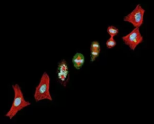
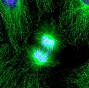
"Cytokinesis: The Dynamic Process of Cell Division Unveiled through Micrographs and Illustrations" Mitosis, a fundamental process in cell division


All Professionally Made to Order for Quick Shipping
"Cytokinesis: The Dynamic Process of Cell Division Unveiled through Micrographs and Illustrations" Mitosis, a fundamental process in cell division, is accompanied by cytokinesis. Through the lens of a light micrograph, we witness the intricate dance of cellular components during cytokinesis. Fluorescent micrographs reveal the mesmerizing beauty as cells divide, showcasing their vibrant fluorescence. Intriguingly, scanning electron micrographs provide us with an up-close view of just-divided HeLa cells. These images capture the precise moment when two daughter cells emerge from their parent cell's embrace. Artwork F006 / 9891 and F006 / 9892 depict this remarkable event with artistic flair. Nuclear division takes center stage in an illustrative masterpiece that showcases its complexity. As plant cells undergo mitosis, a captivating light micrograph captures each step within these green powerhouses. The wonders continue under the gaze of a transmission electron microscope (TEM), revealing dividing cells at an unprecedented level of detail. TEM allows us to explore the intricacies and structural changes occurring during this crucial phase. Even brain cancer cells succumb to cytokinesis; SEM C014 / 0354 exposes dividing brain cancer cells' unique morphology as they multiply relentlessly. Similarly, SEM C017 / 8305 offers insight into HeLa cell division—a testament to their significance in scientific research. Artwork C013 / 4630 delves into lung cancer cell division with striking imagery that conveys both awe-inspiring beauty and somber reflection on this disease's impact on human health. Cytokinesis unravels before our eyes through various imaging techniques—each providing invaluable glimpses into this dynamic process essential for life itself.